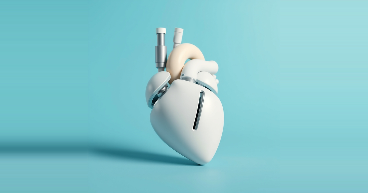
Engineers at UNSW Sydney have built a soft robotic heart that beats, holds simulated blood and contains working mitral valve support structures that can be adjusted to make the valve leak. The device models the left side of the heart and is described in papers in Nature Communications and Advanced Science.
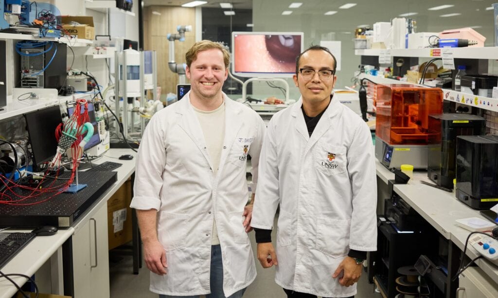
The team is led by Scientia Associate Professor Thanh Nho Do of the School of Biomedical Engineering and the UNSW Medical Robotics Lab. The soft robotic heart is intended as a test bed for diseases and cardiac devices, and as a way to cut early-stage reliance on animal studies.
How the soft robotic heart is built
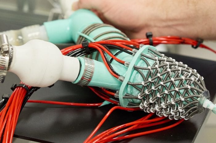
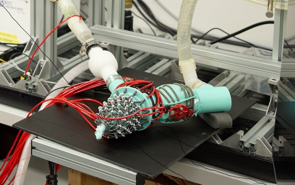
Silicone membranes form the internal chambers of the model. Soft robotic artificial muscles are wrapped around them to reproduce the way the heart contracts and twists. The muscles are arranged to follow the layered muscle architecture of the human heart and are driven by hydraulic pressure, which the team controls to make the ventricular model move.
Dr James Davies, a postdoctoral researcher in Do's group, said the membranes model the inner surface of the human left heart and contain the simulated blood, so the model can pump fluid in and out.
The design includes artificial valves, papillary muscles and chordae tendineae. These structures control the mitral valve, which works like a pair of swinging doors that open and close with each beat. The team says conventional laboratory models do not contain them. Disease can make the valve leaky or stiff, which raises the workload on the heart and contributes to heart failure.
The researchers can change the tension in the artificial papillary muscles. Using that control, they recreated mitral valve prolapse and regurgitation, where blood flows backwards instead of moving efficiently through the heart.
What the tests showed
The team checked the model with ultrasound imaging and with measurements of pressure and blood flow. Invasive pressure and flow measurements were taken in and out of the heart. The model also worked with echocardiography, a non-invasive clinical measure of heart function.
With healthy valve settings, pressure and flow followed physiological expectations. When disease was induced, regurgitation increased and outlet pressure and flow fell, which the researchers say is consistent with human valve disease. Adjusting papillary muscle length changed valve function.
Professor Nigel Lovell, Head of the School of Biomedical Engineering and Director of Tyree IHealthE, said the ultrasound images resembled human cardiac imaging. The team saw human-like valve leaflet motion and blood flow across the valves, including regurgitant jets leaking from valves with induced disease.
A catheter test and a model of HFpEF
The group also placed a newly developed soft robotic cardiac catheter inside the beating model. The catheter moved within the artificial heart and detected when it touched moving cardiac structures. The researchers say this shows how the platform could speed up development of surgical tools.
The model also reproduced changes seen in heart failure with preserved ejection fraction, or HFpEF. Do said the condition accounts for 50 percent of heart failure cases and is among the least well understood and hardest to treat. The team simulated one of its earliest signs, a reduced ability of the heart to relax between beats. Blood then entered the heart more slowly and less efficiently, and the delayed filling raised pressure inside the heart. The team says this closely matches what is commonly seen in patients.
What the team says is still missing
The researchers describe the current model as a proof of concept, not a finished clinical tool. Materials need to improve, control systems need refinement, and the device needs to become more compatible with medical imaging. Future versions must reproduce more aspects of heart function and use patient-specific geometries instead of simplified structures.
The main next step is validation against clinical data. Do said the model must be compared systematically with patient data across a wide range of heart anatomies and disease severities before it can inform clinical decisions. The team also includes Professor Christopher Hayward, a heart failure and transplant cardiologist at St Vincent's Hospital Sydney, along with Professor Jelena Rnjak-Kovacina and Scientia Associate Professor Hoang-Phuong Phan.
The group envisions patient-specific versions built from medical imaging data. Clinicians could use them to compare devices and approaches, and to choose implant type and size, before operating.
Why it matters, and what remains unproven
The notable feature of this soft robotic heart is the valve apparatus. A model that moves like a heart is useful, but one in which the papillary muscles can be tensioned independently lets the same device switch between healthy and diseased valve states. That makes comparisons repeatable. The team's argument for reducing animal use rests on this control: an animal heart cannot be set to a chosen degree of regurgitation on demand.
The catheter test points to a second use. A soft robotic tool that senses contact needs a moving, heart-like environment to be evaluated, and the model supplies one without a living subject. That gives device developers a place to test early designs, though it does not replace later testing.
Several claims remain unproven. The agreement with human physiology so far rests on similar waveforms and imaging features, not on systematic comparison with patient cohorts, and the team says so. The model uses simplified geometry, so results may not transfer to the varied anatomies clinicians see. The patient-specific modelling and surgical planning uses are stated goals, not demonstrated results. Nor has the model been shown to predict how a real implant would perform in a real patient.
The claim that conventional benchtop models cannot reproduce these disease mechanics comes from the team itself. It is plausible given the added valve structures, but it has not been tested head to head in the work described. The next real test of this soft robotic heart is whether independent groups and clinical datasets confirm that its pressure, flow and valve behaviour hold up across many hearts, not one idealised design.